Nano and biology
DNA as a building material
In living cells DNA stores information. In a nanoscience laboratory it can also serve as a construction material: a molecule whose pieces can be programmed to find and bind one another, building shapes that designers draw in advance. DNA nanotechnology is one of the clearest demonstrations of what self-assembly can achieve.
Why DNA
The Wikipedia article on DNA nanotechnology defines the field as the design and manufacture of artificial nucleic acid structures for technological uses, treating nucleic acids as engineering materials rather than as carriers of genetic information. What makes that possible is base pairing. As the article explains, the strict pairing rules mean that only portions of strands with complementary sequences bind together, forming strong, rigid double helices. A designer who controls the sequences therefore controls which strands meet and where, and can in principle specify a target structure with precisely controlled nanoscale features.
DNA has other practical advantages. It can be synthesised to order in any sequence, its double helix is about two nanometres wide and fairly stiff over short lengths, and a century of molecular biology has provided tools to cut, join, copy and analyse it. Few other molecules combine that degree of programmability with such a well-understood toolkit.
Foundations
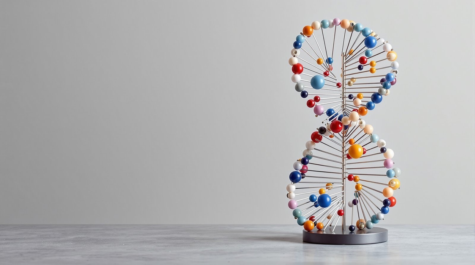
The same article states that the conceptual foundation for the field was laid out by Nadrian Seeman in the early 1980s, and that the field began to attract wide interest in the mid-2000s. Ordinary DNA forms linear double helices, which are not useful for building much. The way around that, used throughout the field, is to design strands that form branched junctions, so that helices meet at defined points and can be joined into larger frameworks; the tetrahedron pictured in the article is built from three-arm junctions at its corners. From those beginnings, the article reports, researchers built two- and three-dimensional crystal lattices, nanotubes, polyhedra and arbitrary shapes, as well as functional devices such as molecular machines and DNA computers.
DNA origami
For two decades most DNA structures were built from many short strands, each designed and purified separately, and the more complex the structure, the harder it became to assemble correctly. In 2006 a different approach appeared. In a paper in Nature, Rothemund described a simple method for folding long, single-stranded DNA into arbitrary two-dimensional shapes. The abstract sets the work in a wider context: bottom-up fabrication is widely used to make relatively simple nanostructures, and a key goal is to reach the complexity routinely achieved by top-down methods.
The method, as the abstract describes it, raster-fills the desired shape with a single-stranded scaffold about 7 kilobases long and chooses over 200 short strands, called staples, to hold the scaffold in place. Once mixed, scaffold and staples self-assemble in a single step. The abstract reports structures roughly 100 nm across that approximate shapes such as squares, disks and five-pointed stars with a spatial resolution of 6 nm. Because each staple can act as a 6-nm pixel, the surfaces can be programmed to carry patterns, and individual structures can be designed to join into larger assemblies, including extended lattices.
The Wikipedia article on DNA origami fills in some background. It notes that the method was developed by Paul Rothemund at the California Institute of Technology, that it was the cover story of Nature on 16 March 2006, and that the scaffold is typically the genome of a bacteriophage called M13. Examples since then include a smiley face, a coarse map of the Americas, and many three-dimensional shapes such as cubes. The article adds that applications have been explored in areas from drug-delivery research to circuitry in plasmonic devices, but that most commercial applications remain at the concept or testing stage.
What DNA structures are used for
The DNA nanotechnology article lists several directions. The structures are beginning to be used as tools in structural biology and biophysics, for example to help determine protein structures by X-ray crystallography and nuclear magnetic resonance. They serve as scaffolds that hold other molecules, nanoparticles or proteins at precisely defined distances, which makes them useful for studying interactions that depend on spacing. And potential applications in molecular-scale electronics and in nanomedicine are, in the article's words, being investigated, which is a fair description of their status: promising research rather than established technology.
Reading DNA nanotechnology papers
Most papers in this field present atomic force or electron microscopy images of the finished structures. The key questions are the same as for any self-assembly paper. What fraction of the product is correctly formed, and how was that measured? How were the imaged structures selected? And do the structures survive the conditions in which they are meant to be used, which may differ greatly from the salt-rich buffer in which they were assembled? A single beautiful image of a folded shape is evidence that the design can work; yield and stability data show whether it usually does.
For the general principles behind DNA assembly, see self-assembly; for the parallel story of peptides, which fold and stack into fibres and tubes, see peptides at the nanoscale; for the machines that DNA and other molecules can form, see molecular machines and nanopores.